
“SKILLED US IMAGING OF THE ADNEXAL MASS”
ILAN E. TIMOR-TRITSCH, MD, AND STEVEN R. GOLDSTEIN, MD
(4 PARTS; SEPTEMBER–DECEMBER 2010)
US imaging made it possible to postpone resection of tumor until delivery
As Dr. Timor-Tritsch and Dr. Goldstein point out, one of the most common adnexal masses is the cyst. In pregnancy, in fact, it is the most common finding.1 Surgical resection is usually reserved for 5- to 10-cm cysts containing septae or nodules, or for cases in which there are solid components.2
I had a 17-year-old patient who presented for prenatal care without ever having had a gynecologic exam. The only significant aspect of her history was use of dextroamphetamine and amphetamine (Adderall) for attention deficit hyperactivity disorder during the early first trimester. Once her self-test for pregnancy yielded a positive result, however, she stopped taking the medication.
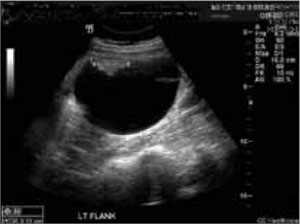
FIGURE 1 Imaging at 9 weeks of gestation
Teratoma or solid component of the collision tumor.At 7 weeks’ gestation, I performed her first pelvic exam, during which I palpated a mass in the left adnexa. The patient was asymptomatic. US imaging revealed a complex cystic mass measuring 4.7 × 4.4 × 3.7 cm on the left ovary and a viable 9-week gestation in the uterus (FIGURE 1). A repeat US at 18 weeks revealed that the mass had increased in size to 7.5 × 6.9 × 3.7 cm (FIGURE 2). The patient (and her mother) refused resection, so a follow-up scan was scheduled. The patient remained asymptomatic.
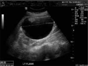
FIGURE 2 US scan at 18 weeks
Serous cystadenoma or cystic component of the collision tumor.Follow-up scans were performed over the remainder of the pregnancy, and the mass eventually reached 13.3 cm at its greatest dimension. At 28 weeks, the family again refused resection, opting to wait until term. The patient was placed on bed rest. Primary cesarean delivery was performed at term because the fetus was unengaged. The mass was resected without any complications to infant or mother (FIGURE 3). The infant was a viable male with Apgar scores of 8 and 9, and mother and baby were discharged after 72 hours.
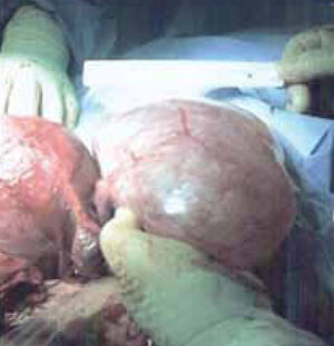
FIGURE 3 Delivery
Gross anatomy of the collision tumor.As for the cyst, it was not malignant but was a mature cystic teratoma with a benign serous cystadenoma—otherwise known as a collision tumor. A collision tumor represents the coexistence of two adjacent but histologically distinct tumors.3
Even though a cesarean delivery had to be performed in this case, the outcome was good.
Solid and complex ovarian tumors that have solid components discovered during pregnancy generally should be treated surgically because of the low but significant risk of cancer (1%–10%).4 But it’s good to know that it is possible to follow a tumor to term in a compliant patient. In this case, US was of great value; without it, surgery could not have been postponed until term.
The most frequent and serious complication of a benign ovarian cyst during pregnancy is torsion, which is fairly common in the first trimester. And if a large cyst remains in the pelvis until labor, it may block the outlet and lead to uterine rupture.2 Most experts agree that elective resection is warranted for an ovarian cyst larger than 6 cm that persists after 16 weeks’ gestation.
Joseph DeRosa, DO
Ironton, Ohio
We want to hear from you! Tell us what you think.